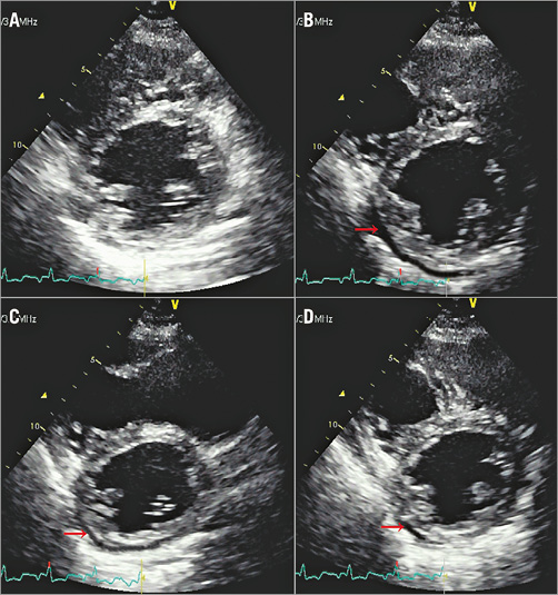

The prognosis of viral pericarditis is generally good, with a self-limited course, and if uncomplicated, patients may be treated on an outpatient basis. Patients often experience a prodrome of an upper respiratory tract infection. Viral PericarditisĬoxsackievirus B and echovirus are the most common viruses, and a fourfold increase in antiviral titers is required for the diagnosis. Most cases are presumed to have a viral etiology. The cause of acute pericarditis is often difficult to establish, and idiopathic pericarditis remains the most common diagnosis. The rub may disappear with the development of an effusion and impending cardiac tamponade. It is best heard during inspiration at the left lower sternal border, with the patient leaning forward. This is felt to be caused by fibrinous deposits in the inflamed pericardial space ( Figure 2) the timing of which can be mono-, bi-, or triphasic (corresponding to atrial systole, ventricular systole, and early ventricular diastole, respectively). Positional changes are characteristic with worsening of the pain in the supine position and with inspiration and improvement with sitting upright and leaning forward.Ĭlassically, a scratchy, grating, high-pitched friction rub (which has been likened to the squeak of leather of a new saddle) is heard. Metastatic: lung, breast, bone, lymphoma, melanomaĪcute pericarditis typically presents with acute onset severe, sharp retrosternal chest pain, often radiating to the neck, shoulders, or back.Post-Myocardial infarction (early and late) 1 These are discussed in more detail later.īox 1: Common Causes of Pericarditis and Pericardial Effusion Other common causes include infection, renal failure, myocardial infarction (MI), post-cardiac injury syndrome, malignancy, radiation, and trauma. The most common form of acute pericarditis is idiopathic, which accounts for about 90% of cases ( Box 1). It occurs more commonly in men than in women. PrevalenceĪcute pericarditis is the admitting diagnosis in 0.1% of hospital admissions and 5% of admissions with chest pain. 2 Generally, the diagnosis requires 2 of these 3 features. 1 Acute Pericarditis DefinitionĪcute pericarditis is an inflammatory process involving the pericardium that results in a clinical syndrome characterized by chest pain, pericardial friction rub, changes in the electrocardiogram (ECG) and occasionally, a pericardial effusion. While the pericardium is not critical for life it serves important functions including maintenance of cardiac position within the chest and as a barrier to infection and inflammation. Between the two layers of the serous pericardium lies the pericardial cavity which normally contains up to 50 mL of pericardial fluid ( Figure 1).

The visceral pericardium or epicardium is closely adherent to the underlying myocardium and is reflected upon itself to form the outer parietal pericardium which lines the fibrous sac.


The inner portion of the pericardium is a two-layered sac called the serous pericardium. The outer layer of the pericardium is referred to as the fibrous pericardium and usually measures less than 2 mm in thickness. The pericardium is a thin fibroelastic sac composed of two layers that separate the heart from the surrounding mediastinal structures.


 0 kommentar(er)
